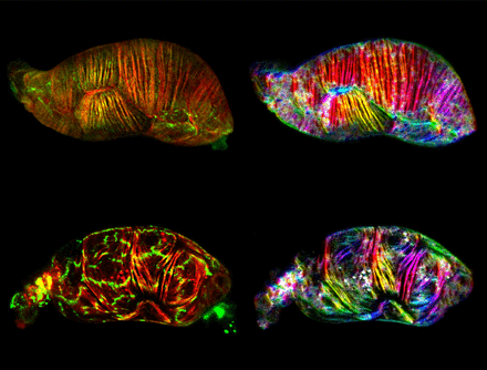
Caenorhabditis elegans (C. elegans) is a unique organism that allow for observation of their physiological processes just by looking under a microscope – because their bodies are clear! This characteristic has provided PhD student Alison Wirshing great potential into understanding and visualizing the reproductive process as an egg moves into the uterus of this nematode. Her observations and techniques led to the discovery of spermathecal cell organization that occurs at the moment of ovulation.
Her research, in collaboration with the Cram Lab of the Department of Biology, was published in the Molecular Biology of the Cell this July, with her discoveries featured as the cover photo (see above image).
The Cram Lab studies how cells receive and react to mechanical stressors. Wirshing, in collaboration with Professor Erin Cram, has been studying how the cells in the reproductive system of the nematode Caenorhabditis elegans (C. elegans) coordinate stretching and contracting actions that allow for an egg to move through reproductive organs in order to become fertilized and develop.
C. elegans are hermaphrodites, carrying both egg and sperm. Their transparent structure allows for visibility of all internal processes, like their self-reproduction. One worm produces 300 eggs (oocytes) throughout its lifetime, and each oocyte follows a specifically coordinated process in the worm before hatching. Developing oocytes grow in the gonad arm before cells contract to push oocytes into the spermatheca. The spermatheca is where fertilization occurs. Once fertilized, spermathecal contractions will push the egg into the uterus.
The cells of the spermatheca contain actin and myosin bundles, proteins that work together to form networks of contractile fibers, allowing for the structure to stretch and contract, in this case to push the egg into the uterus. It had been previously observed that these cells, before and after ovulation, look very different. Before ovulation, the fibers appeared as a disorganized mesh of actin bundles. However afterwards, all actin bundles in each cell appear organized in the same direction. What was making this happen? Enter Wirshing.
She used confocal microscopy to create 4D images of the spermatheca stretching and contracting to push an egg through, in order to be able to visualize the exact moment when actin organization occurs. Through a combination of fluorescent protein labels and other visualization tools and data analysis, Wirshing was able to determine that the peak of actin organization occurs at the moment when the cell is contracting to push the egg forward.
These results open up the possibility of understanding how other cells respond to mechanical stress. The actin cytoskeleton and many of the regulatory proteins Wirshing discovered occur in many species’ systems, including medically important systems like the human bronchial airways.
“One of the cool things to me is these two types of actin networks; basal and apical. They are on different surfaces, have different networks, and it would be so exciting to determine the role of each regulatory protein and what it does in its network,” Wirshing said.
Wirshing is also interested in visualizing force landscapes – how mechanical stresses vary across the whole spermathecal tissue – to determine the relationships between forces and actin orientation.
Both Wirshing and Cram are very excited about this paper because they were able to determine so much about cell contractility, occurring as close to its natural state as possible, and now there is great potential for more exciting research to follow.

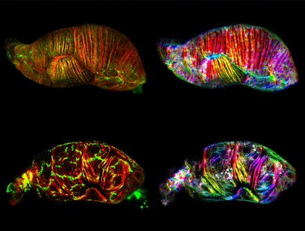
Visualization of the C. elegans spermatheca, the organ that pushes eggs into the uterus. On the left in red and green are actin (the cells’ “skeleton”) and myosin (the “motor” to contract cells) respectively. On the right, fibers are color coded based on their angle of orientation. (Image courtesy of Alison C. E. Wirshing and Erin J. Cram, Department of Biology, Northeastern University)

