

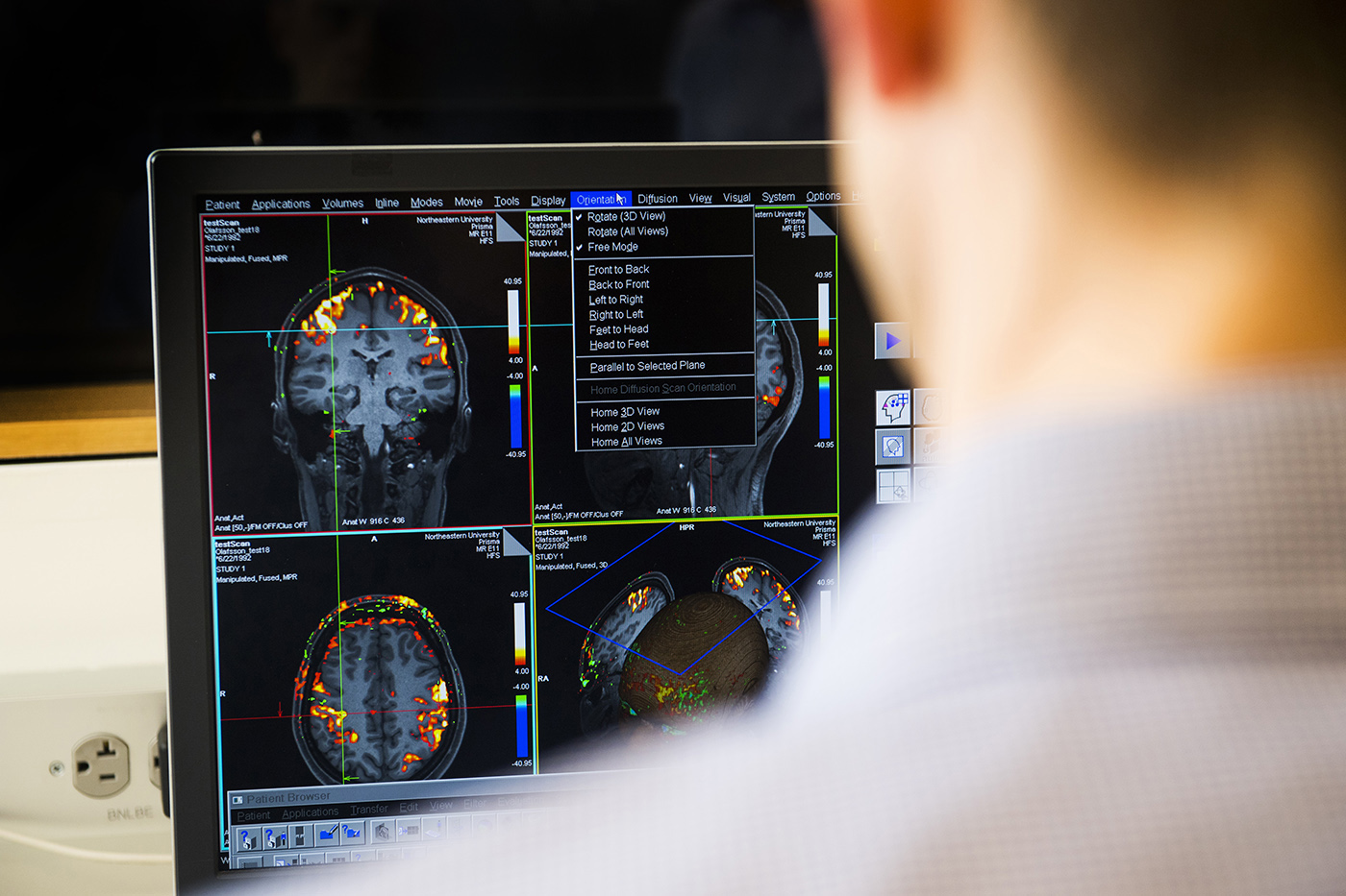
Your brain is always working. Even as you’re lying still, thinking of nothing in particular, blood is rushing oxygen to different areas of your brain, creating a network of activity. Through brain imaging, researchers can watch this resting activity rise and fall in different regions. Some areas will fluctuate together, like pairs of dancers moving to the same beat.
These patterns of synchronized activity form what researchers refer to as resting state networks, said Susan Whitfield-Gabrieli, a neuroscientist at Northeastern. “It has turned out that these are very informative and can predict things like clinical outcome.”
Resting state networks can vary from person to person. In a study published earlier this month, Whitfield-Gabrieli and her colleagues found that one particular pattern may be an early sign of schizophrenia.
Susan Whitfield-Gabrieli has found that a particular pattern of brain activity may be an early sign of schizophrenia. Photo by Adam Glanzman/Northeastern University
“It’s a different organization of the brain that we can actually see,” said Whitfield-Gabrieli, a psychology professor who directs the biomedical imaging center at Northeastern. “If you have this type of connectivity, you are three times more likely to develop schizophrenia.”
People who have schizophrenia can experience hallucinations and delusions, struggle to keep their thoughts in order or express emotions, and suffer from other cognitive impairments. They’re typically diagnosed between their late teens and early 20s, but early symptoms, such as disordered thinking or difficulty focusing, can start several years before the first psychotic episode.
The study focused on 158 patients at the Shanghai Mental Health Center who had experienced early symptoms and were considered “high risk” for developing schizophrenia, as well as 93 individuals with no signs of psychosis. The researchers took brain images of all of them and then tracked their progress.
Over the next year, 23 of the high-risk individuals were diagnosed with schizophrenia after some sort of psychotic episode. Their resting state networks, imaged at the beginning of the study, were noticeably different from the other high-risk individuals and the control group.
“You see a distinct correlation pattern,” Whitfield-Gabrieli said.
The region of the brain responsible for processing sounds, known as the superior temporal gyrus, is typically coupled with the areas of the brain that handle sensory inputs and responses. In the individuals that developed schizophrenia, however, this region was linked to the limbic regions of the brain, which process emotions.
“Auditory hallucinations is one of the cardinal symptoms of schizophrenia,” Whitfield-Gabrieli said. This connection may help explain why those hallucinations occur, or at least what makes them so powerful.
These links already existed before the patients’ first psychotic episodes. But that doesn’t necessarily mean that the patients were born with them. The connections in our brain change as we grow up. Whitfield-Gabrieli hopes to use these imaging techniques on younger children to identify the first signs of schizophrenia, and perhaps change the course of the disease.
“There are many types of interventions that, if it’s detected early enough, might be very helpful,” Whitfield-Gabrieli said. “They may even prevent developing the disorder.”
This story was originally published by News@Northeastern on November 26, 2018.
