
We already have all sorts of maps – of highways and hiking trails, of floor plans and tunnels, even of skeletal systems and the evolution of our ancestors. Now, for the first time, we will have a blood volume map of the human brain – a detailed layout of the complicated network of vessels that make up our circulatory system for arguably the most important organ in our body.
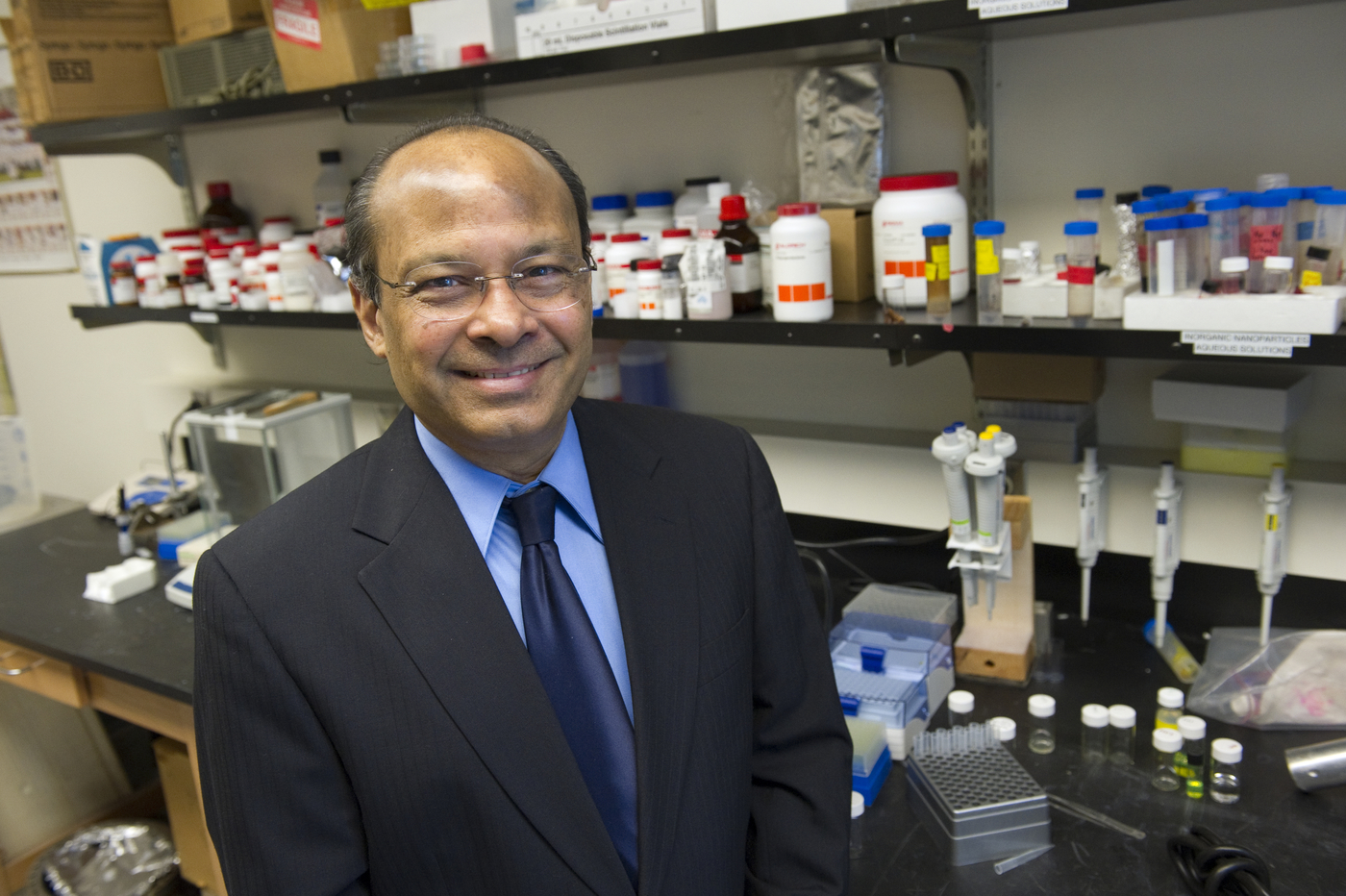
A new MRI brain imaging technique developed by Northeastern researcher Srinivas Sridhar will use magnetic nanoparticles to create the first atlas of blood volume in the human brain. The results will be used to better identify and address neurological diseases, such as Alzheimer’s and drug addiction. A groundbreaking clinical trial is already underway at Massachusetts General Hospital (MGH).
The technique, called QUTE-CE MRI, or Quantitative Ultra-short TE Contrast-Enhanced MRI, was developed three years ago by University Distinguished Professor of Physics, Bioengineering, and Chemical Engineering Srinivas Sridhar and his students, Codi Gharagouzloo, Ju Qiao, and Liam Timms.
The technique utilizes iron oxide nanoparticles which attract to the blood flowing through the body. The nanoparticles appear bright under the MRI, creating extremely detailed images of every small and large vessel in the bloodstream. The use of iron oxide is a newer, safer technique for MRI imaging that is increasing in popularity.
Sridhar wanted this technique to have an immediate impact on people, so he created a company called Theranano LLC, which translates the Northeastern discovery into technology that can be used in the clinic. Through a National Institute of Health/National Institute on Drug Abuse (NIH/NIDA) funded grant, Theranano, in joint partnership with Northeastern, will be testing the QUTE-CE MRI technique at MGH to create the first clinical brain images of cardiovascular networks.
“It’s really exciting, and it’s what I’ve wanted to do my whole life. I write lots of papers, but I want something that helps and directly impacts people. That’s really why this is so exciting, because we made the discovery at Northeastern, and already within just a couple of years, we’re in the clinic,” Sridhar said.
The produced images will be the start of an entirely new class of MRI imaging. The results will be able to provide information on where blood is in the brain and any anomalies that could be related to diseases and issues. The trial will consist of two parts. First will be to create a snapshot of all brain vasculature, giving a picture of the blood flow in the brain and compiling the first brain atlas of cerebral blood volume. This atlas will outline the amounts of blood in each part of the brain, which will be an important template to compare to in cases where blood flow in the brain isn’t working properly.
The second part of the trial consists of comparing MRIs of normal subjects to those who have any sort of an anomaly or disease. This includes patients with neurological diseases such as Alzheimer’s and Parkinson’s, traumatic brain injuries, as well as drug addictions.
“This is the first time I’m going to test in a clinic – and we are excited to see what the results will be, we really don’t even know yet. We know what the imaging looks like in animals and everyone agrees those are spectacular,” Sridhar said. “But what’s most satisfying is to see my idea, developed with the help of my students, now already in the clinic to test in humans and study neurological diseases.”
The trial has already begun its first steps at MGH and will be accepting its first patients very soon. The connection between industry and academia helps Northeastern embody its initiative of use-inspired research that has an important purpose and will impact the community.
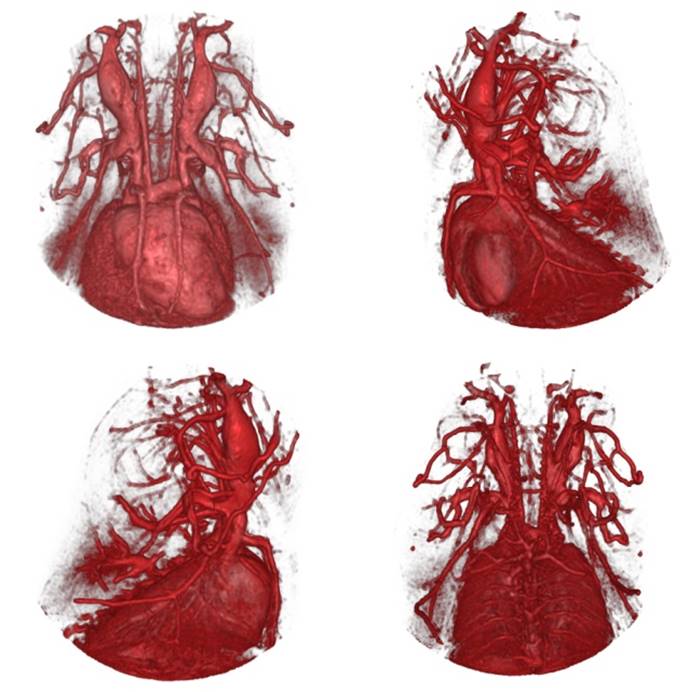
QUTE-CE MRI enables images of rat vasculature with unprecedented clarity and definition. Photo courtesy of Srinivas Sridhar
