The Barry L. Karger Medal in Bioanalytical Chemistry
Established by the Barnett Institute of Chemical and Biological Analysis at Northeastern University in honor of its founding director, Dr. Barry L. Karger, the Karger Medal is an annual award recognizing an individual who has significantly contributed to the development of new bioanalytical methods and high impact in the life sciences.
Barry L. Karger Medal in Bioanalytical Chemistry Celebration
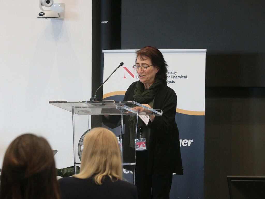
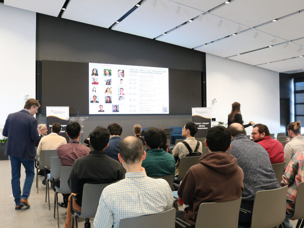
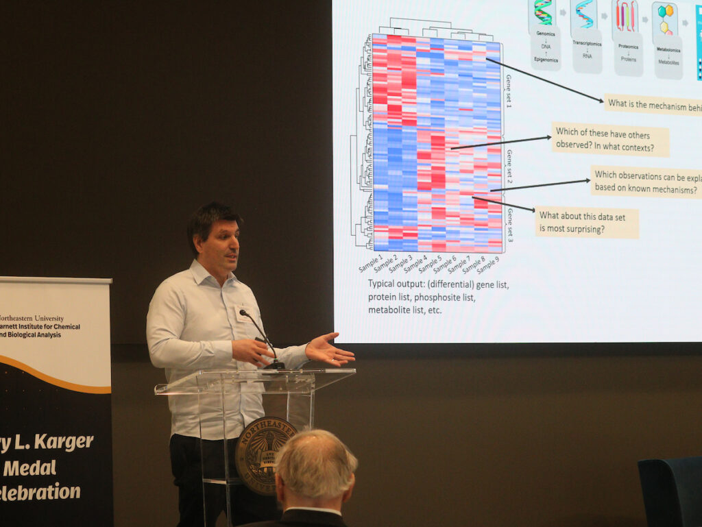
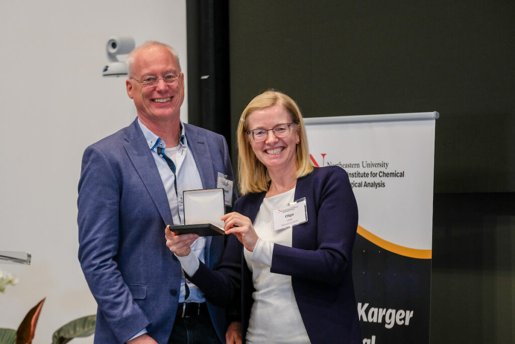
The 2026 Barry L. Karger Medal in Bioanalytical Chemistry Celebration took place on March 12 at Northeastern University. This prestigious award recognizes groundbreaking advancements in biopharmaceutical characterization, proteomics, and systems biology technologies. We were delighted to present the 2026 Karger Medal to Dr. Ron Heeren, a renowned expert in mass spectrometry-based molecular imaging.
2026 Karger Medal Recipient
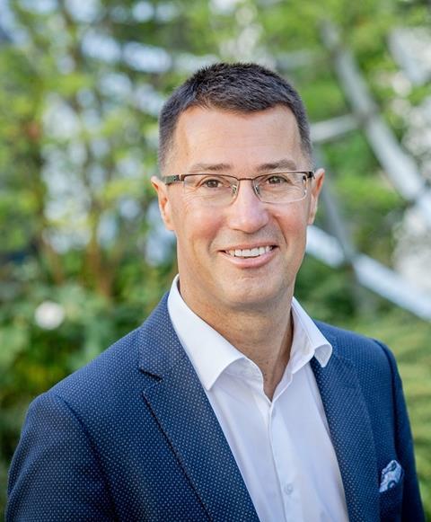
Ron Heeren, PhD
Distinguished Professor and Limburg co-chair of the Maastricht Multimodal Molecular Imaging Institute
Professor Heeren is internationally recognized for his pioneering work in mass spectrometry–based molecular imaging. His research has transformed how we visualize and understand molecular patterns in health and disease, enabling insights from the molecular level to clinical applications in pathology and surgery.
By advancing mass spectrometry–based chemical microscopy, his work bridges physics, chemistry, biology, and medicine to reveal how local tissue environments influence molecular signaling. These innovations are helping pave the way toward predictive, personalized medicine and tissue regeneration, bringing high-resolution molecular imaging closer to routine clinical practice.
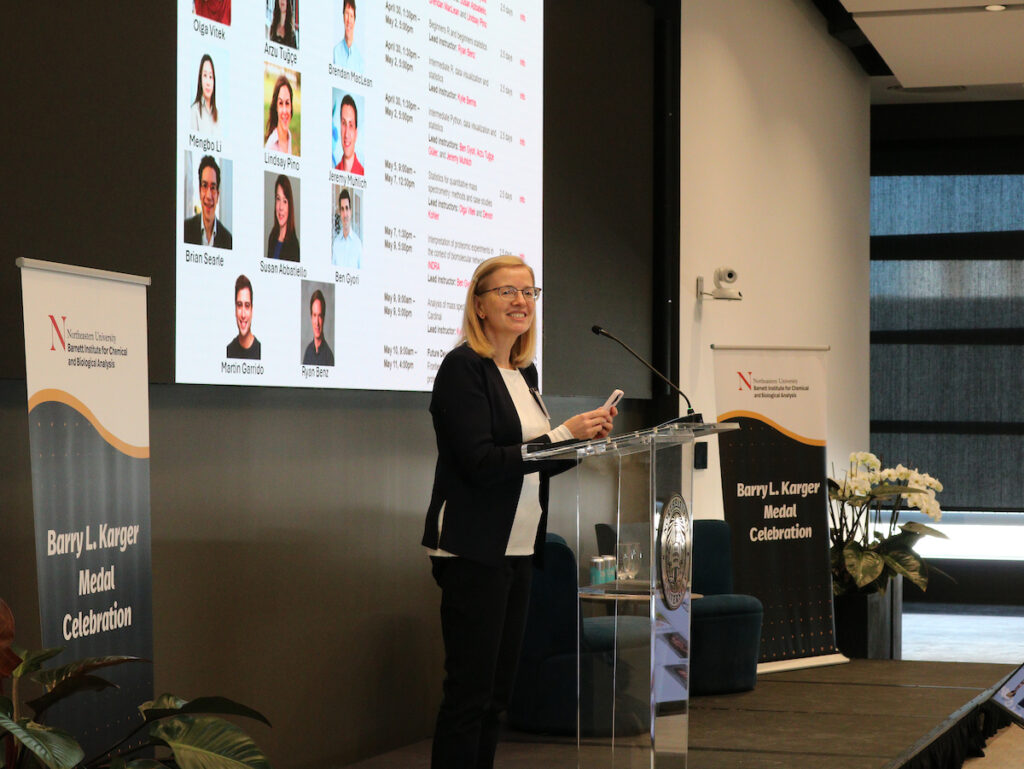
Featured Speakers
-
Ron Heeren, PhD
Karger Medal Awardee
Founding Director, Maastricht Multimodal Molecular Imaging Institute -
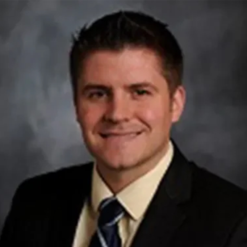
Brian Hoffman, PhD
Director, Protein Sciences and Mass Spectrometry Services, The Jackson Laboratory
Adjunct Faculty, Medical College of Wisconsin -
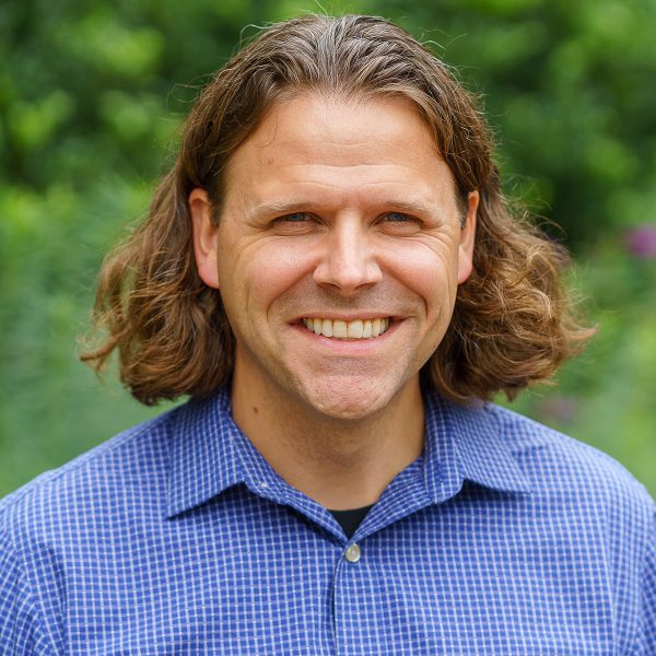
James Monaghan, PhD
Professor, Biology, Northeastern University
Collaborator, Barnett Institute for Chemical and Biological Analysis -
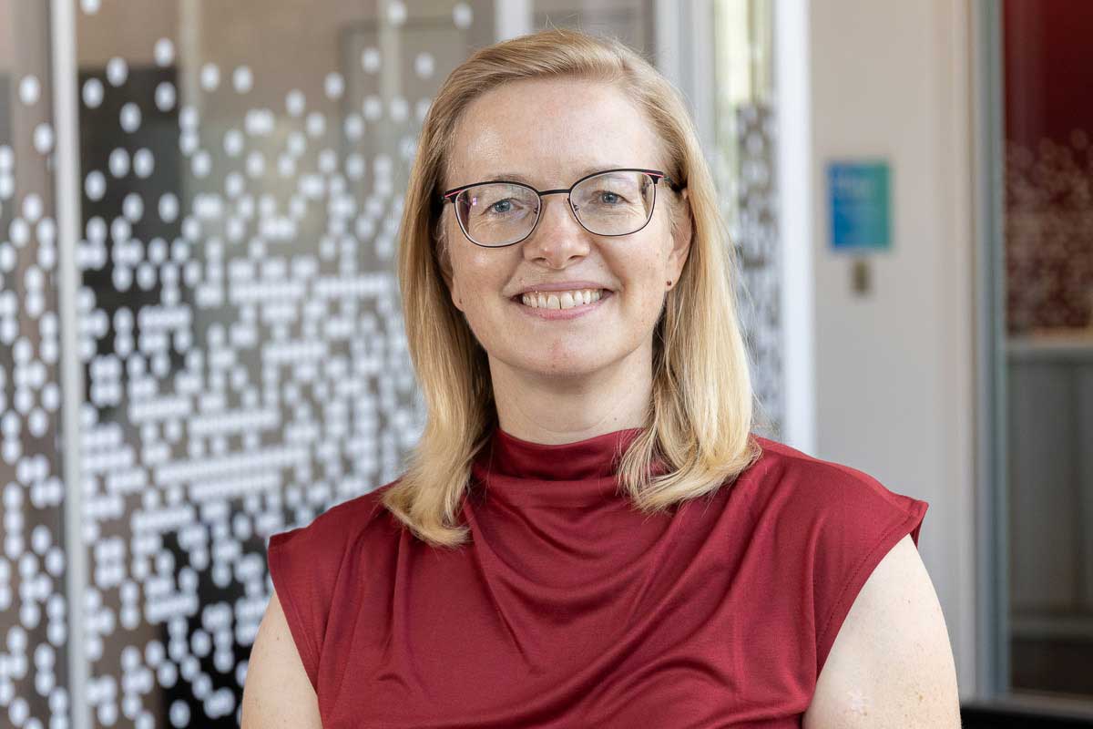
Olga Vitek, PhD
Director, Barnett Institute for Chemical and Biological Analysis
Professor, Khoury College of Computer Sciences
Northeastern University -

Brent Nelson, PhD
Interim Dean, College of Science
Professor, Physics
Northeastern University
Abstracts
Ron Heeren, PhD
Imaging Complexity: Visualizing Nature with Bioanalytical Chemistry
The understanding of complex biological processes in their spatial context is crucial to modern molecular medicine and requires innovations in bioanalytical technologies. Mass Spectrometry based spatial biology is revolutionizing fundamental and applied life science research. This implies that future diagnostics will no longer rely on a single molecule but require insight into the intricate biochemical interplay of molecular pathways across the omics.
As such, bioanalytical chemistry technologies in the “omics” arena play an increasingly important role in unraveling life’s complexity. Technological advances have increased methodological sensitivity, allowing researchers to acquire detailed molecular information of smaller and smaller samples imperative to understand cellular or subcellular chemistry. The biggest challenge is to put that concerted information in the context of the biological problem the samples originate from.
As a result, innovative molecular imaging technologies at the single cell level, have impacted both fundamental and translational biomedical research. Sensitive and selective molecular microscopes in modern spatial biology offer new insights in spatial and molecular complexity of cellular metabolism that contextualize cellular function in health and disease.
Imaging mass spectrometry has evolved to demonstrate it’s ability to visualize this almost inconceivable complexity. It’s capabilities of revealing contextual, local molecular information on the microscale aid researchers to provide fundamental insights into biological processes that will impact the future of personalized health and care.
Brian Hoffman, PhD
Multi-Omics Integration to Define the Molecular Architecture of the Aging Brain
Brian Hoffmann is the Director of Protein Sciences and Mass Spectrometry Services at The Jackson Laboratory, where his lab utilizes state-of the-art mass spectrometry technologies to advance a variety of research areas, including research on the aging brain and Alzheimer’s Disease (AD).
His presentation will focus on his collaborative research utilizing a combination of spatial lipidomics, proteomics, and transcriptomics to identify molecular mechanisms in the brain. Through these high-resolution multi-scale approaches, they are able to investigate protein and lipid homeostasis in the aging and AD brain at the single cell level.
In addition, the region of interest information collected from the spatial omics approaches is utilized to inform the specific neurons that are targeted for patch-proteomics experiments in which electrophysiology is coupled single neuron proteomes. This approach was utilized to define single cell proteomic changes in aging and presymptomatic neurons of wild-type and AD mouse models across ages to identify molecular changes before onset of the symptomatic disease state.
James Monaghan, PhD
Deciphering The Cellular and Molecular Basis of Axolotl Limb Regeneration Using Multimodal Imaging and Omics
The axolotl salamander (Ambystoma mexicanum) is one of the few vertebrates capable of regenerating complex structures, including an entire adult limb, following amputation. Unlike mammals, which respond to injury with scar formation, axolotls mount a highly coordinated regenerative response in which the remaining tissues are remodeled, and a new limb is rebuilt with remarkable fidelity. Understanding how this process is orchestrated across dozens of cell types through dynamic tissue reorganization demands approaches that can resolve molecular and cellular identity within their spatial tissue context.
This presentation highlights our use of single-cell transcriptomics, multiplexed RNA imaging, whole-mount tissue clearing, genetic reporter animals, and functional gene perturbation to map the cellular landscape of the regenerating limb. Through this integrative approach, we have identified candidate signaling pathways that mediate intercellular communication and are functionally required for regeneration to proceed.
These findings illustrate how multimodal imaging and omics strategies can unlock the complexity of regenerating tissues, with broader implications for understanding tissue repair across vertebrates.
Olga Vitek, PhD
Statistics, Machine Learning and AI for Interpreting Mass Spectrometry Imaging Experiments
Mass spectrometry imaging (MSI) characterizes the spatial distribution and molecular composition of tissues at high resolution. This technology is broadly applied in biological and clinical research, including disease diagnosis, prognosis, and drug delivery studies. Our lab aims to enable robust statistical analyses of high-dimensional MSI experiments with complex designs, improve interpretability of MSI datasets, and facilitate reproducible workflows for these applications. The open-source package Cardinal supports advanced functionalities for data preprocessing, for interpretation of MSI experiments with statistical, machine learning and AI algorithms, and for efficient handling of large datasets through out-of-memory data structures.
This presentation overviews the functionalities that we have recently added to Cardinal and its companion software packages. These include TIMSImaging for processing trapped ion mobility mass spectrometry imaging data; functionalities for statistical analysis of differentially abundant analytes in experiments with multiple biological replicates, conditions, and regions of interest; and Teardrop for co-registration of images from multiple complementary biomolecular modalities.
We demonstrate using several case studies that these developments increase the scope of analyses available to the research community through free and open source software, and hope that this will facilitate even broader adoption of MSI.
2025 Karger Medal Highlights
Speakers and Abstracts
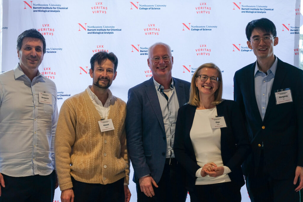
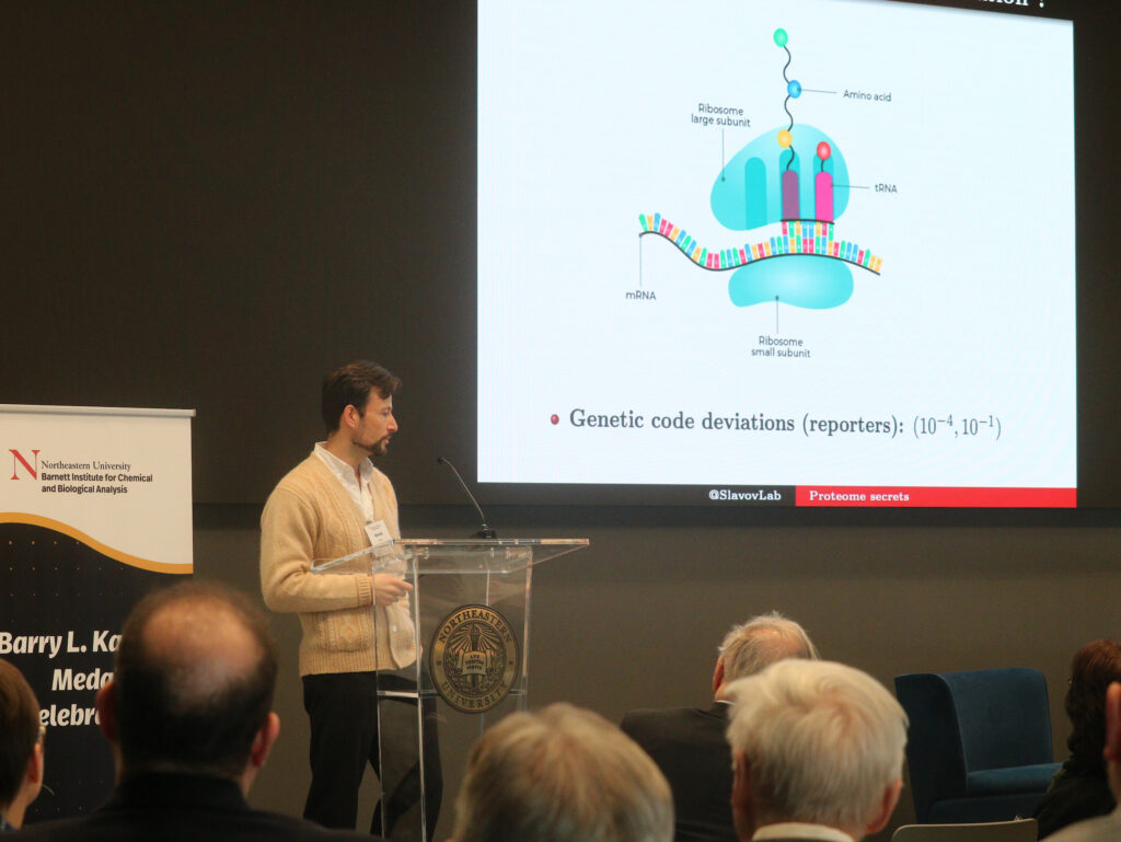
Nikolai Slavov, PhD
Professor, College of Engineering, and Faculty Fellow, Barnett Institute for Chemical and Biological Analysis, Northeastern University
From Protein Variation to Biological Functions
Biological functions are reflected in the natural variation of proteome configurations across individual cells. Single-cell proteomics methods may decode this variation and empower inference of biological mechanisms with minimal assumptions. This promise is beginning to be realized by sensitive and scalable mass spectrometry methods. I will discuss approaches that have allowed us to measure and interpret protein covariation in different biological systems, including primary macrophages and melanoma cells expressing markers for drug-resistance priming. The focus of the talk will be on conceptual innovations and data interpretation leading towards molecular mechanisms.
Benjamin Gyori, PhD
Associate Professor, Khoury College of Computer Sciences and the College of Engineering, and Faculty Fellow, Barnett Institute for Chemical and Biological Analysis, Northeastern University
Large Scale AI-Assisted Knowledge Integration for Protein Biology
Discovery in biomedicine requires interpreting experiments in the context of prior knowledge and existing data on the function of proteins and other biomolecules. However, this process is fundamentally limited by the fact that the large body of existing data and published knowledge is fragmented such that it cannot be readily used in an actionable form by scientists.
Through examples from our recent work, we introduce the key components of a technical framework that could overcome these issues. First artificial intelligence-based data annotation and machine reading, coupled to semantic technologies leveraging bio-ontologies enable large-scale data integration across literature and structured databases.
From this unified knowledge base, predictive and explanatory models of protein function and cellular behavior can be assembled that allow interpreting novel experimental observations and proposing hypotheses to be validated experimentally.
These technologies enable a cycle between experimentation and data interpretation in which human scientists can leverage machine-assisted data integration and interpretation to accelerate discovery.
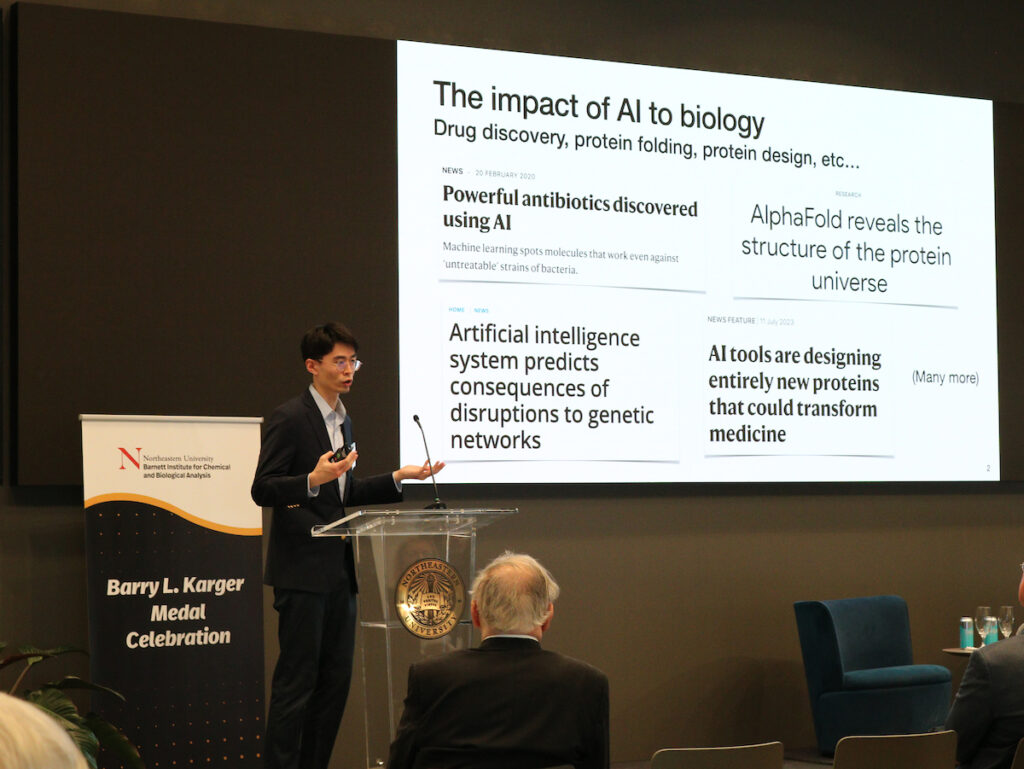
Wengong Jin, PhD
Assistant Professor, Khoury College of Computer Sciences and Faculty Fellow, Barnett Institute for Chemical and Biological Analysis Northeastern University
Accelerating Drug Discovery Via Geometric and Generative AI
AI for drug discovery is an emerging field that aims to computationally design new proteins or molecules with desired properties. Traditional experimental approaches to drug discovery are time-consuming and labor-intensive, due to the large combinatorial search space of molecule and protein structures.
In this talk, I will describe how to accelerate drug discovery via novel generative and geometric deep learning methods. First, I will introduce junction tree variational autoencoder (JT-VAE), a generative model for molecular graphs. Inspired by probabilistic graphical models, JT-VAE leverages the low tree-width of molecular graphs and represents a molecule as a junction tree of chemical motifs.
Second, I will present Neural Euler’s Rotation Equation (NERE), an equivariant rotation prediction network inspired by rigid-body dynamics. Based on NERE, they developed an unsupervised binding energy prediction method that estimates the likelihood of a protein complex via SE(3) denoising score matching.
Lastly, I will demonstrate how these algorithmic innovations make real-world impacts on drug discovery. Through collaboration with biologists in wet labs, they successfully designed new antibiotics to fight against antimicrobial resistance and new antibodies with potential for cancer immunotherapy.
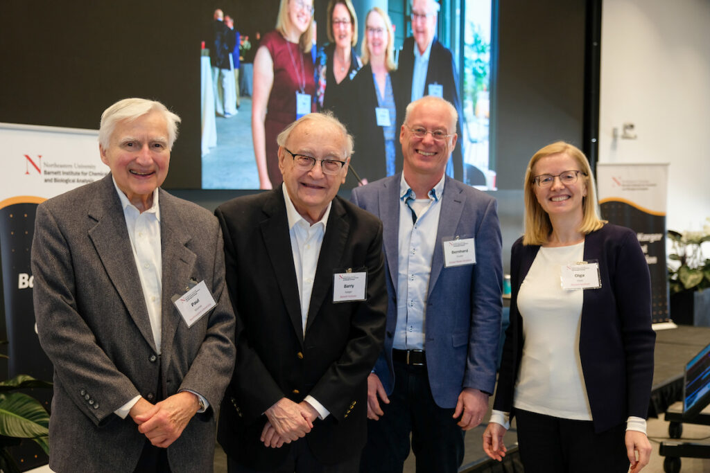
Bernhard Küster, PhD
Professor of Proteomics and Bioanalytics at Technical University of Munich and Director of the Bavarian Biomolecular Mass Spectrometry Center
Understanding What Therapeutic Drugs Really Do
Almost all drugs act on proteins, are proteins, make or degrade proteins and it has been known since the days of Paracelsus that drugs exert their effects in a dose-dependent fashion. The molecular processes leading to a drug-induced change in cellular phenotype can be roughly divided into: i) target binding, ii) pathway engagement, and iii) cellular reprogramming to arrive at a new viable state or cell death, together forming the mechanism of action (MoA) of a drug. Today, quantitative mass spectrometry is the most comprehensive approach for the proteome-wide characterization of drugs on all three levels because of its unique ability to assay thousands of proteins and their post-translational modifications in complex cellular backgrounds in parallel.
In this presentation, I will introduce the proteome-wide decryptT, decryptM and decryptE technologies that measure target deconvolution, pathway engagement and cellular reprogramming in a fully dose-dependent fashion respectively. Based on the analysis of >3,000 drugs including small molecules and antibodies, examples for drug characterization at all three levels will be discussed, particularly focusing on unexpected or even surprising findings. These include drug repurposing opportunities for kinase inhibitors, the long elusive MoA or Rituximab or the loss of T-cell receptor components in T-cells in response to HDAC inhibitors.
We have developed CurveCurator to put proteome-wide dose-response measurements on a solid statistical foundation and deposited the millions of dose-response curves and derived cellular EC50 values obtained into proteomicsdb.org for FAIR data sharing and mining by the scientific community. Examples for how the data may be used will be highlighted by ascribing new functions to proteins and phosphorylation sites based on their consistent responses to drugs that target known proteins, signaling pathways or cellular machines. We expect that the various implementations of the general “decrypt” approach will become a standard in drug discovery and pharmacology.
Past Recipients

Dr. Bernhard Küster
Professor of Proteomics and Bioanalytics, Technical University of Munich
Director, Bavarian Biomolecular Mass Spectrometry Center
Lecture: Understanding What Therapeutic Drugs Really Do
Bio
Professor Küster conducts research in the fields of chemical proteomics and precision medicine. Together with an interdisciplinary team of biochemists, molecular biologists and bioinformaticians, he deals with questions about how exactly therapeutic drugs work, which molecular mechanisms play a role in cancer and how these can be used for individual approaches in clinical treatment.
After studying chemistry at the University of Cologne, Professor Küster completed his doctorate in biochemistry at the University of Oxford. He then worked as a postdoctoral researcher in Heidelberg and Odense, Denmark. Before taking up a full professorship at TUM in 2007, he was Vice President of Cellzome (now GSK). Professor Küster is Director of the Bavarian Biomolecular Mass Spectrometry Center and Co-Director of the Center for Infection Prevention. Professor Küster is co-founder of the biotech companies OmicScouts and MSAID.
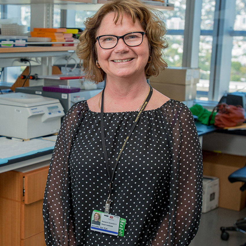
Dr. Jenny van Eyk
Professor and Director, Basic Science Research
Barbra Streisand Women’s Heart Center Cardiology, Cedars-Sinai
Lecture: The two sides of precision medicine: Proteomic enablement of biomarkers and therapeutics
Bio
Jennifer Van Eyk, PhD, is an international leader in the area of clinical proteomics and her lab has focused on developing technical pipelines for de novo discovery and larger scale quantitative mass spectrometry methods. This includes multiple reaction monitoring (MRM, also known as SRM) and most recently data independent acquisition.
Dr. Van Eyk’s laboratory is well known for the extreme technical quality of the data generated, rigorous quality control with tight %CV while applying these to key clinical questions. The aim is to maximize throughput and reproducibility in order to move targeted and robust discovery methods into large population healthy continuous assessment and clinical grade assays focusing on brain and cardiovascular diseases.
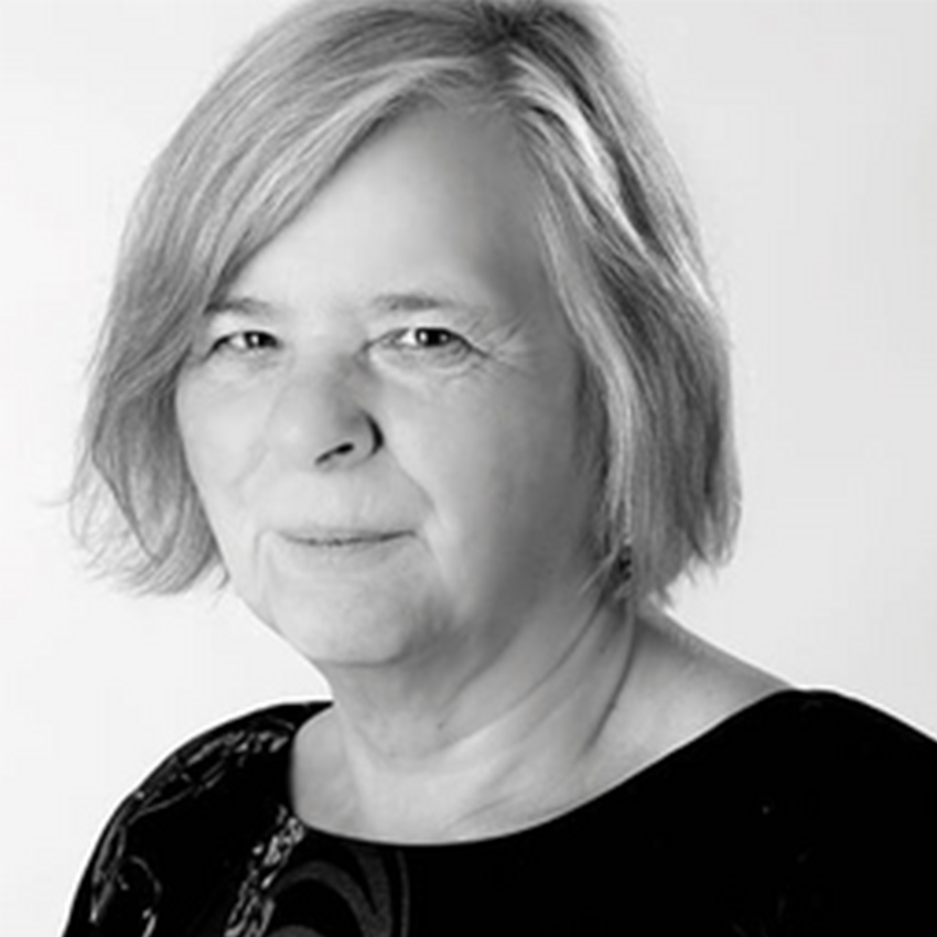
Dr. Pauline Rudd
Emeritus Fellow the Conway Institute and Visiting Investigator
Bioprocessing Technology Institute, AStar, Singapore
Lecture: Advances in glycomics and its critical role in systems biology
Bio
Pauline Rudd is a research professor of glycobiology at University College Dublin. She heads the GlycoSciences Research Group at the National Institute for BioProcessing Research and Training in Ireland (NIBRT) where she is a PI and consultant. She has more than 280 scientific publications and has given over 350 lectures and seminars at international meetings. Before moving her group to Dublin in 2006, Professor Rudd was a member of the Oxford Glycobiology Institute for 25 years, where she was a Senior Research Fellow and a University Reader in Glycobiology at the University of Oxford.
Professor Rudd obtained a BSc in chemistry at the University of London and a PhD in glycobiology at the Open University, UK. She was a Founding Scientist of Wessex Biochemicals (later Sigma London), Visiting Research Associate at The Scripps Research Institute, CA, Visiting Professor of Biochemistry at Shanghai Medical University PRC, Visiting Scientist at Ben Gurion University of the Negev, Israel and an Erskine Visiting Fellow, Canterbury University, Christchurch, New Zealand. She is a Fellow of the Royal Society of Medicine, London, visiting Professor at St. George’s Hospital, London and an Adjunct Professor at North Eastern University, Boston, NUI Galway, Trinity College Dublin and at University College, Dublin. She is a VI at BTI, AStar, Singapore. In 2014 she was awarded an Honorary Doctorate at Gothenburg University, Sweden.
In addition to her basic research interests, she has many links with pharmaceutical companies across the world because GlycoScience is a major area of specialised expertise required to ensure the safety and efficacy of biotherapeutic drugs, such as monoclonal antibodies for cancer and autoimmune disorders. In 2010 Prof Rudd was awarded the James Gregory Medal and an Agilent Thought Leader award and in 2012 she received a Waters Global Innovation award. In 2014 she was awarded an Honorary Doctorate at Gothenburg University, Sweden.
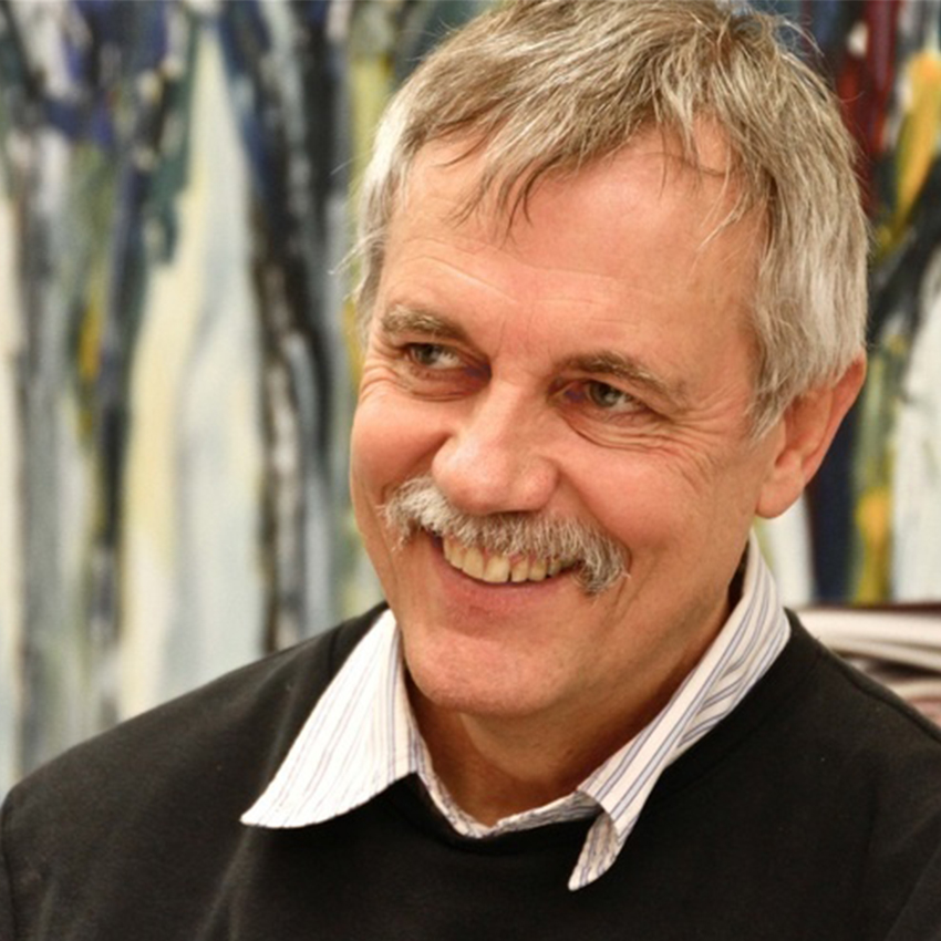
Dr. Rudolph Aebersold
Head, Department of Biology
Institute of Molecular System Biology ETH Zurich, Zurich, Switzerland
Lecture: SWATH-MS: Principles, current state and new developments
Bio
Ruedi Aebersold is one of the pioneers in the field of proteomics. He is known for developing a series of methods that have found wide application in analytical protein chemistry and proteomics like a new class of reagents termed Isotope Coded Affinity Tag (ICAT) reagents used in quantitative mass spectrometry. Professor Aebersold and his team of researchers use the protein profiles determined by this method to differentiate cells in different states, such as noncancerous versus cancerous cells, and to systematically study how cells respond to external stimuli. These “snapshot” profiles indicate which cells contain abnormal levels of certain proteins. This is expected to lead to new diagnostic markers for disease and to a more complete understanding of the biochemical processes that control and constitute cell physiology.
Professor Aebersold serves on the Scientific Advisory Committees of numerous academic and private sector research organizations and is a member of several editorial boards in the fields of protein science, genomics, and proteomics.
He is a native of Switzerland and obtained his PhD in Cellular Biology at the Biocenter of the University of Basel in 1983. Since that time, he is a faculty member of the Universities of Washington and British Columbia, until 2000, when he co-founded the Institute for Systems Biology in Seattle. In 2004, he accepted a position as full professor at the Institute of Biotechnology at the Swiss Federal Institute of Technology (ETH) in Zurich, where in January 2005, his research group became the first integral part of the newly founded Institute of Molecular Systems Biology.

Dr. Matthias Mann
Director, Department of Proteomics and Signal Transduction
Max Planck Institute of Biochemistry, Munich, Germany
Lecture: High Resolution LC MS/MS for cell signaling and biomedical research
Bio
Matthias Mann, a scientist in the area of mass spectrometry and proteomics, is the inaugural recipient of the Barry L. Karger Medal in Bioanalytical Chemistry.
Born in Germany, Dr. Mann studied mathematics and physics at the University of Göttingen and obtained his PhD in chemical engineering at Yale University. After a postdoctoral fellowship at the University of Southern Denmark in Odense he became group leader at the European Molecular Biology Laboratory (EMBL) in Heidelberg. Later he went back to Odense as a professor of bioinformatics and in 2005 took up a director position at the Max Planck Institute of Biochemistry in Munich. Additionally, Dr. Mann was appointed the director of the proteomics department of the Novo Nordisk Foundation Center for Protein Research in Copenhagen.