

 by Greg St. Martin
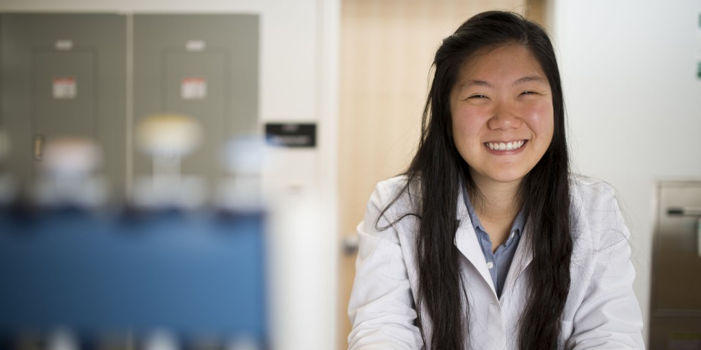
Ellie Shin was determined to find a co-op working in a wet lab on campus.
She enrolled at Northeastern as a history major but took science courses during her freshman year, setting her sights on a pre-dental track to explore her passion for working in healthcare. After her first year, Shin inquired about co-op opportunities with some of the university’s newest faculty members and caught the attention of Jon Tilly, the new chair of the Department of Biology. Tilly arrived at Northeastern in July of 2013 and together with Dori Woods, an assistant professor in the Department of Biology, established the Laboratory for Aging and Infertility Research (also called the LAIR) as a cutting-edge and multi-investigator research space.
Shin started working with Tilly and Woods in the LAIR as a volunteer, turned that opportunity into a co-op from July to December 2014, and has continued working there through a directed study this semester. The third-year student has also bolstered her academic resumé, adding a biology major and a mathematics minor.
The LAIR focuses on women’s health, with a particular interest in infertility and healthy aging. Tilly and Woods have produced groundbreaking research on the application of regenerative medicine in reproductive biology, leading to several issued patents that are currently in clinical study.
“It’s been very exciting,” Shin, SSH/S’16, said of working in the lab. “Professors Tilly and Woods have given me a lot of responsibility in designing my own experiments and writing my own protocols.”
Shin’s work focuses on two projects, one being a collaborative effort with other LAIR team members that builds upon Tilly and Woods’ research on mitochondria, which are tiny structures within all cells that perform a range of important tasks. “Without mitochondria,” Tilly said, “we wouldn’t be here.”
Tilly and Woods want to better understand mitochondria and their regenerative abilities, particularly because of their important implications for both fertility and healthy aging. Knowing that when mitochondria respire they make energy, they developed a novel process to separate out single mitochondria by attaching probes that fluoresce when this energy is made. Shin, for her part, developed a new method for using a scanning electron microscope to take a 3-D image of a mitochondrion, which is about one micrometer in diameter.
“I was in charge of visualizing mitochondria using a technique called scanning electron microscopy,” Shin explained. “The more conventional technique is TEM (transmission electron microscopy), where you’re cutting the sample into slices. But these are only two-dimensional images, and we felt SEM would offer more robust images of the samples we’re working with and a better look at the characteristics of the mitochondria we’re collecting.”
This is a very tedious technique that requires a great deal of trial and error, but in December Shin produced the group’s first high-quality images. Prior to her efforts, many scientists who study mitochondria questioned if what Shin accomplished was even technically feasible.
Shin’s other project, which she has worked on independently in the LAIR, involves replicating the protocols and findings of Mariusz Ratajczak of the University of Louisville, whose team several years ago discovered new very small embryonic-like cells in bone marrow that have significant regenerative properties apart from creating blood cells. Ratajczak reported that these VSELcells were pluripotent, meaning that they can give rise to any cell type. This is important, Shin said, because they could potentially be used as an alternative to human embryonic stem cells in research and medicine, which is controversial.
But several notable scientists have refuted Ratajczak’s work, so Shin aims to help settle an important debate in stem cell biology: whether these VSEL cells exist. She is presenting her work to date on this project at a poster session during the 2015 Experimental Biology conference in Boston on March 28–April 1.
Both research projects require a high-tech piece of equipment that enables fluorescence activating cell sorting, or FACS. Here’s how it works: you feed a mix of cells or organelles, each type of which is labeled with a distinct fluorescent probe, into the machine. Next, the machine marches them single-file down a tube. Lasers then read each one and individually sort those that fluoresce the same, so you can separate out and study the ones you want. The rest are collected separately for comparative analysis.
Tilly and Woods specially engineered the FACS machine in the LAIR to enable detection of a single mitochondrion, and Shin has quickly become the resident expert. “It’s been such a joy to watch her learn and grow in the lab,” Tilly said. “She’s now a gifted FACS technician.”
Shin earned the annual Schafer Medical Research Scholarship, which is named after distinguished alumnus Andrew Schafer, LA’69, and supports students’ co-op experiences conducting laboratory medical research under the guidance of a faculty mentor for six-months. Students also receive mentorship from Schafer, who is professor of medicine in hematology-oncology and director of the Richard T. Silver Center for Myeloproliferative Neoplasms at Weill Cornell Medical College.
Originally published in news@Northeastern on March 31, 2015.
